Flat Panel Detector C-Arm Machine
SHOC-CS01 Medical Imaging
This product has been discontinued and removed from our shelves.
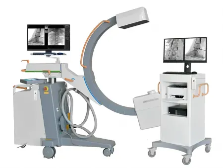
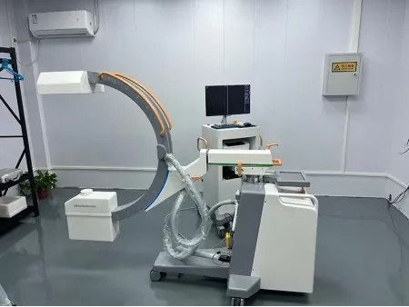
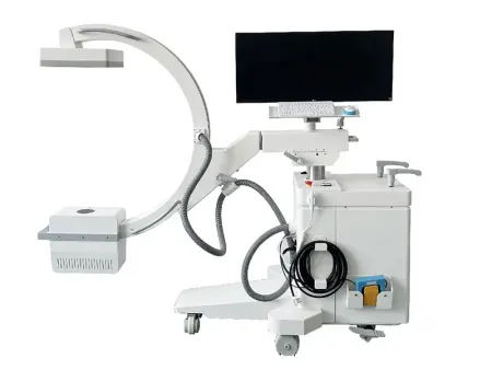
The flat panel detector C-arm machine gives you exceptional imaging quality with an easy-to-use design, making it a valuable tool for precise and efficient diagnostics. With its C-arm structure, you get multi-angle imaging, high-resolution diagnostics, and smooth operation, helping you make accurate clinical decisions faster.
Designed to enhance your workflow, this system features a high-frequency generator that delivers high-quality, high-penetration X-rays for clear, detailed imaging. The 21 cm × 21 cm flat panel detector is larger than traditional image intensifiers and uses amorphous silicon technology, giving you higher density and spatial resolution for improved surgical accuracy.
For long-duration procedures, the oil-cooled, heat-resistant X-ray tube ensures consistent image acquisition, while the 34-inch monitor allows you to view real-time video and reference images at the same time, speeding up your diagnostic process. The microcomputer control system includes self-diagnosis and automatic protection, with a dual-CPU design that improves system stability and safety.
With a large open design, this system gives you more freedom during procedures. It supports handheld, footswitch, and remote control operation, letting you make adjustments easily from inside or outside the operating room. Plus, with DICOM 3.0 compatibility, you can easily send, receive, and print images via PACS.
This detachable C-arm machine is a popular choice in the market, offering exceptional imaging quality with its French Trixell flat panel detector and Italian IMD technology X-ray tube. The high-performance amorphous silicon detector ensures clear, detailed imaging, making it a reliable tool for medical professionals.
The 21 × 21 cm image sensor, featuring directly deposited cesium, provides high-quality low-dose imaging with a frame rate of up to 30 frames per second, delivering sharp, real-time visuals.
For those seeking advanced functionality, an optional motorized system allows for fully automated C-arm movements, improving efficiency and ease of use. This system is widely used in orthopedics, neurosurgery, pain management, and emergency departments, enhancing diagnostic accuracy and procedural outcomes.
HF generator
120 kV, 5 kW, 40 kHz
- Fluoroscopy mode 40-120 kv 0.5-5 mA (kV manual and automatic);
- Pulsed fluoroscopy 40-120 kV 0, 5-5 mA
- Boost fluoroscopy 40-120 kV 5-10 mA;
- Photography 40-120 kV 1-250mAs
- Dual-focus Rotating anode: 0.3 mm / 0.6 mm, 100 mA
- Thermal capacity: 800kHU
- Italy IMD Technology
- France Trixell brand
- Plate: single A-Si TFT photodiode plate
- Scintillator: Csl
- Total transmitted image width: 1024 (1344 optional)
- Total transmitted image height: 1024 (1344 optional)
- Pixel pitch: 200 um
- DQE: 80%
- Quantization depth: 16
- Power supply input: 24±10%
- Communication/image transfer: Ethernet cable
- Cold start feature
- Offset stability validity up to 10 minutes
- Fluoroscopy and RAD images capability
- Passive cooling device
- High speed image transfer using Ethernet link
- 5 Degree recursive Noise Reduction
- Frame freezing
- Thousands of image data storage
- Image conversing and rotating
- Screen display with image comparison
- Support “one click” print, print the image report
- Equip with Dicom 3.0 network port
- Support Dicom 3.0standard data export function
- DAP (optional)
- Vertical travel: 0-400 mm(electric), Horizontal travel: 0-200 mm (electric optional)
- Rotation about horizontal axis: ±180°(electric optional )
- Slipping along the circular arch rail (electric optional)
- Distance of focal spot to image generator(SID):1000 mm
- Depth in arm ≥ 650 mm Mobile stand: 1800×800×1850mm
- Image system: 750×530×1680 mm
- Flat panel detector: 1set
- X-ray generator: 1set
- Working station 24'' Dell LCD high definition display 1 set
- Working station 24'' Dell LCD high definition display 1 set
- Mobile stand:1 set
- Inkjet Printer: 1set
- Handset wired exposure brake: 1set
- Wireless handset exposure brake: 1set
- Foot switch for fluoroscopy: 1set
If you need clear, high-resolution imaging with low radiation exposure, this flat panel C-arm machine is the perfect choice for orthopedics, neurosurgery, pain management, and emergency procedures.
With its high-performance amorphous silicon detector, you get exceptional image quality and precise diagnostics. The 21 × 21 cm image sensor with directly deposited cesium (CsI) provides low-dose imaging with a frame rate of up to 30 frames per second, giving you sharp, real-time visuals for surgical precision.
Designed to improve your workflow and patient care, this system integrates seamlessly into mobile C-arm fluoroscopy X-ray units, making it an ideal solution for vascular and surgical applications.
- Ambient temperature: 10℃ to 40℃
- Relative humidity: 30-75%
- Atmospheric: 70-106 kPa
| Model | SHOC-5KW | SHOC-15KW |
| Output power (kW) | 5 | 15 |
| Current of fluoroscopy (mA) | 0.3-6.3 | 0.3-6.3 |
| Current of radiography (mA) | 0.1-100 | 0.5-150 |
| Pulsed mA (mA) | 32 | 32 |
| Voltage (kV) | 40-125 | 40-125 |
| mAs range (mAs) | 0.2-100 | 0.2-100 |
| ms range (ms) | 10-1600 | 10-1600 |
- Anode type: 5kw is Stationary Anode tube, 15kW is Rotating Anode tube
- Focal Spot value:0.3 mm / 0.6 mm
- Rotating anode speed:2800 rmp
- Anode heat storage:300 kHU
- Housing Heat Storage Capacity:1200 kHU
- Size:21×21 cm
- Detector Technology: Amorphous Silicon
- Scintillator:CsI
- Image resolution:2.5 lp/mm
- Frame rate:Max. 30 frames/sec
- Active pixels:1024×1024 pixels
- A/D conversion:16 bit
- Pixel Pitch: 205 μm
- Faster image capture speed:The image will be displayed on the monitors in 0.8s after exposure.
- Brand:JPI
- Ratios:10:1
- Density:60l / cm
- SID: 100 cm
- SID:1000 mm
- Free space(Tube to I.I):820 mm
- Arc depth:640 mm
- Vertical movement:0-400 mm (Electric)
- Horizontal movement:0-200 mm (Manual)
- Planning movement:±15°(Manual)
- Orbital movement: -30° to 90° (Manual)
- C-arc rotation:±180°
- Monitor:One set 34’’ LCD LG brand HD monitor
- Computer memory:500 G mass storage.
- CPU:2.7 GHZ
- Software:The image can be step-less rotated continuously for 360°.Flip the Reference image horizontally or vertically. Applies various Gamma values for each image Last frame frozen Images can be saved in different formats, such as JPG, DCM, RAW, etc. DICOM 3.0, Data communication, Data storage etc.


