Ceiling-Mounted X-Ray Machine
SHO-CMX01-New Medical Imaging
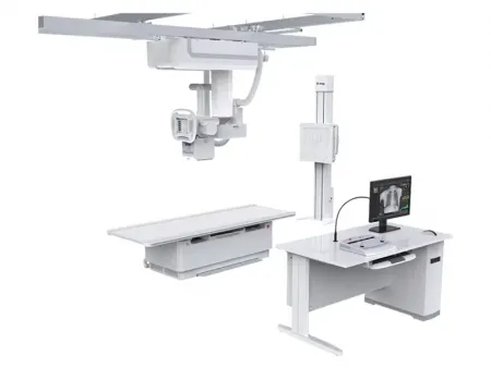
The SHO-CMX01-New ceiling-mounted digital radiography machine gives you reliable, high-quality imaging for chest, limbs, abdomen, and lumbar spine exams. Whether you're working in a radiology department, medical center, diagnostic center, health examination center, orthopedics, or trauma unit, this machine is designed to support all your radiographic needs.You’ll appreciate its ability to handle conventional imaging, special radiography with image stitching, precise visual radiography, large-area fracture radiography, and comprehensive physical examination imaging. It’s built to serve every clinical department in your general radiology setup.
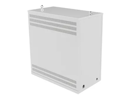
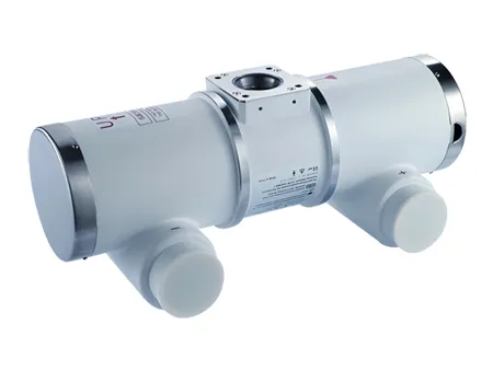
Your machine comes equipped with a powerful 65.5kW generator, a trusted imported Canon high-speed X-ray tube for consistent, long-term use, and dual flat panel detectors (wired and wireless) that give you ultra-clear images. The motorized electric collimator helps you quickly adjust the beam, saving you time with each exam.
You’ll also find the 10.4-inch large touchscreen control panel and workstation with monitor, computer, joystick controller, foot brake, and image processing software make your daily work smooth and efficient. Everything is designed so you can focus on your patients and deliver accurate, dependable diagnostics every time.
| Item | Content | Technical Parameter |
| Power | Voltage | 380 V |
| Frequency | 50 Hz±1Hz | |
| Capacity | 105 kVA | |
| Internal Resistance | ≤ 0.17 Ω | |
| X-Ray Generator | Power | 65.5 kW |
| Inverter Frequency | 500 kHz±20% | |
| Radiography Tube voltage | 40 kV - 150 kV | |
| Radiography Tube current | 10 mA - 80 0mA | |
| Radiography Exposure time | 1.0~10000 ms | |
| Radiography mAs | 0.1 mAs - 800 mAs | |
| Collimator | Brightness | >100 Lux |
| Visible light illumination | 5 s ~ 45 s (every step 5s). | |
| Filter | ≥1 mmAL | |
| X-Ray Tube | Model: | E7254FX |
| Tube focus: big/small | 1.2 mm / 0.6 mm | |
| Input power | 180 Hz: Big focus 102 kW Small focus 40 kW | |
| Anode thermal capacity | 285kJ (400 kHU) | |
| Rotary anode speed | 9700 rpm (180Hz) | |
| Component thermal capacity | 950 kJ (1339 kHU) | |
| Target angle | 12° | |
| Fixed filter | 0.8 mm Al / 75 kV | |
| Flat Panel Detector*2 PLD1717X PLD1717V3 | Active area | 427 (H) mm × 427(V) mm |
| Pixel pitch | 139 μm | |
| Pixel matrix | 3072 (H) ×3072 (V) | |
| Limiting resolution | Unattenuated body mode ≥ 3.7 lp/mm 2 5mm thick aluminium attenuated body mode ≥ 3.4 lp/mm | |
| A / D transition | 16 bit | |
| Acquisition speed | Up to 30 fps | |
| Energy range | 40 - 150 kVp | |
| Computer Workstation | 19inch monitor computer keyboard mouse speaker | |
| Joystick remote controller | ||
| Foot brake control system | ||
| Desk | ||
| Image processing software | ||
- Registration: Includes regular registration, emergency registration, adding agreements, adding items, clearing information, and starting inspections.
- Work list: View list information, search for patients to be examined, refresh the to-be-examined list, delete examinations, and adjust display column settings. Start inspection or emergency registration as needed.
- Exam list: View list information, display and search examined patient data, delete images, store images, burn CDs, add items, adjust display column settings, and modify examination information.
- Patient size options: Thin adults, adults, and larger adults.
- Photography parameter settings: Adjust exposure mode, frame rate setting, kVp, mA, ms, mAs, AEC, and focus selection.
- Perspective parameter settings: Configure exposure mode, frame rate setting, kVp, mA, ABS, and time reset.
- Browsing tools: Zoom, flip horizontal, flip vertical, rotate left 90 degrees, rotate right 90 degrees, zoom in, zoom out, return to original size, move image, invert color, adaptive size, ROI, magnifier, default window width and window level, ROI window width and window level, visible window width and window level, point gray value, advanced processing, and ellipse gray measurement.
- Measurement tools: Arrow, cardiothoracic ratio (CTR), distance measurement, angle measurement, and spine measurement
- Canon high-speed X-ray tube.
- Suitable for long-time, high-intensity exposure.
- High rotating speed for fast heat dissipation and extended service life.
- 500 kHz ultra-high inverter frequency, 800 mA maximum tube current, stable radiation output, excellent radiation quality, and good imaging effect.
- High-power, high-voltage generator ensures stable, high-quality radiation output.
- Guaranteed after-sales service and low maintenance costs.
- Two FPDs supporting both wired and wireless radiography functions.
- 17” x 17” ultra-high-definition pixel FPD for a larger field of view, eliminating the need to move the detector to observe the entire dynamic process.
- Advanced and efficient flat panel technology delivers distortion-free images with high resolution, providing an accurate basis for clinical diagnosis.
- During visualization or replay, if suspected lesions are found, millisecond high-definition spot shots can capture single-frame images at any time, allowing doctors to further diagnose, reduce misdiagnosis, and quickly compile reports.
- Quickly select and preset the desired field of view, saving time during patient positioning.
- According to shooting needs or technician habits, a single key switches the beam range. The conventional beam limiter only switches between two radiography beam ranges, while the automatic electric collimator adjusts beam size intelligently based on body position, greatly improving inspection efficiency.
- Newly added touchable table-side screen with multifunctional features.
- Synchronized with the station, allowing radiographers to create new exams in both the X-ray room and control room for greater convenience in daily exams and emergencies without repeating settings.
- Preview images after exposure.
- Real-time display of SID stretching.
- Set exposure parameters easily.
- Select body parts for exposure using human graphic illustrations.
- Adjust the collimator-effective area.
- Digital display of tube head rotation angle.
- 3D model positioning demonstration.
- Gravity sensor with automatic rotation.
- Emergency exam access: automatically records radiography first and completes patient registration later when urgent diagnosis is needed.
Looking for a ceiling-mounted X-ray system that enhances efficiency and accuracy? Contact us today to find the right solution for your facility!


